Home

razen Sklenite zavarovanje hipoteza colors of fluorescence markers microscopy Izmerljivo orkester striženje

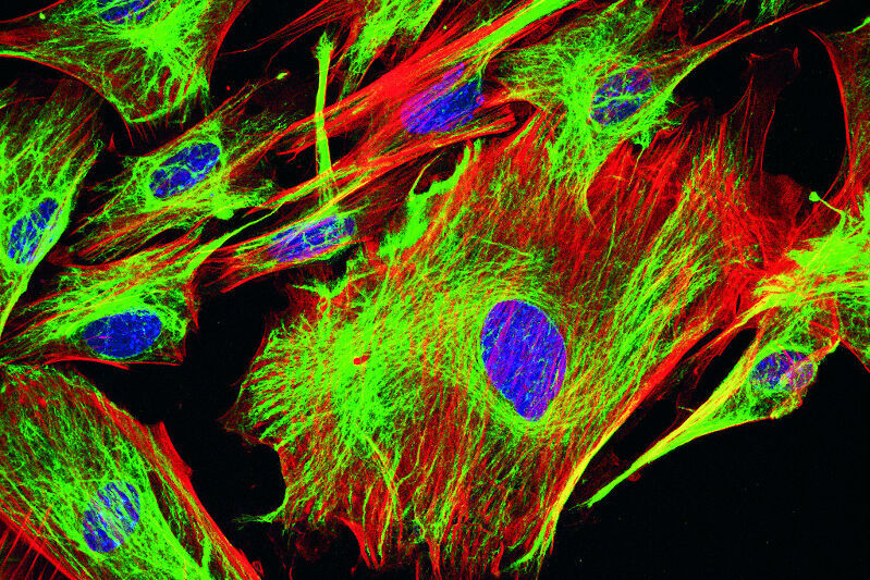
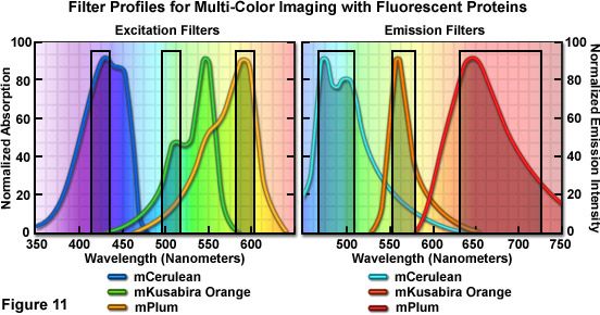
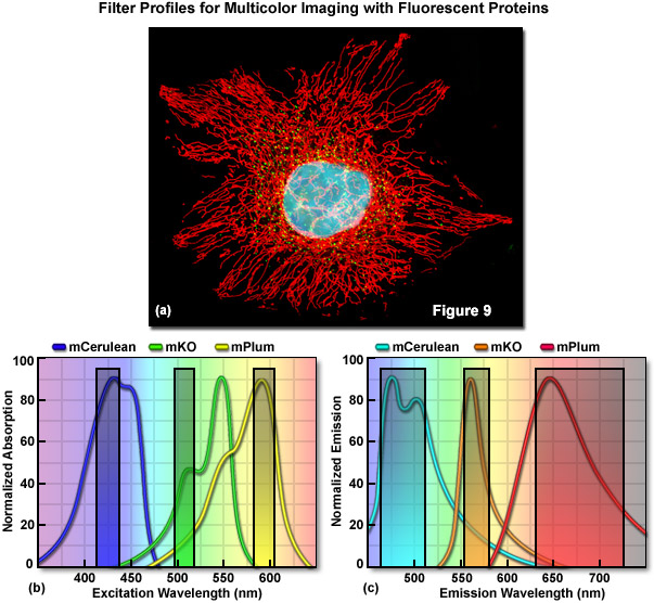
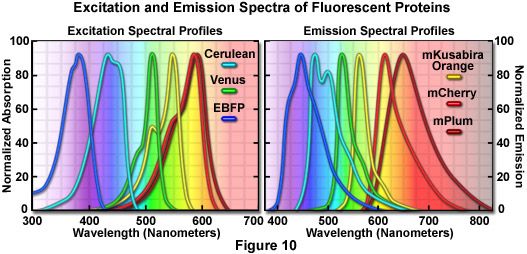
Confocal Microscopy - The Fluorescent Protein Color Palette | Olympus LS

Fluorescent Dyes | Science Lab | Leica Microsystems

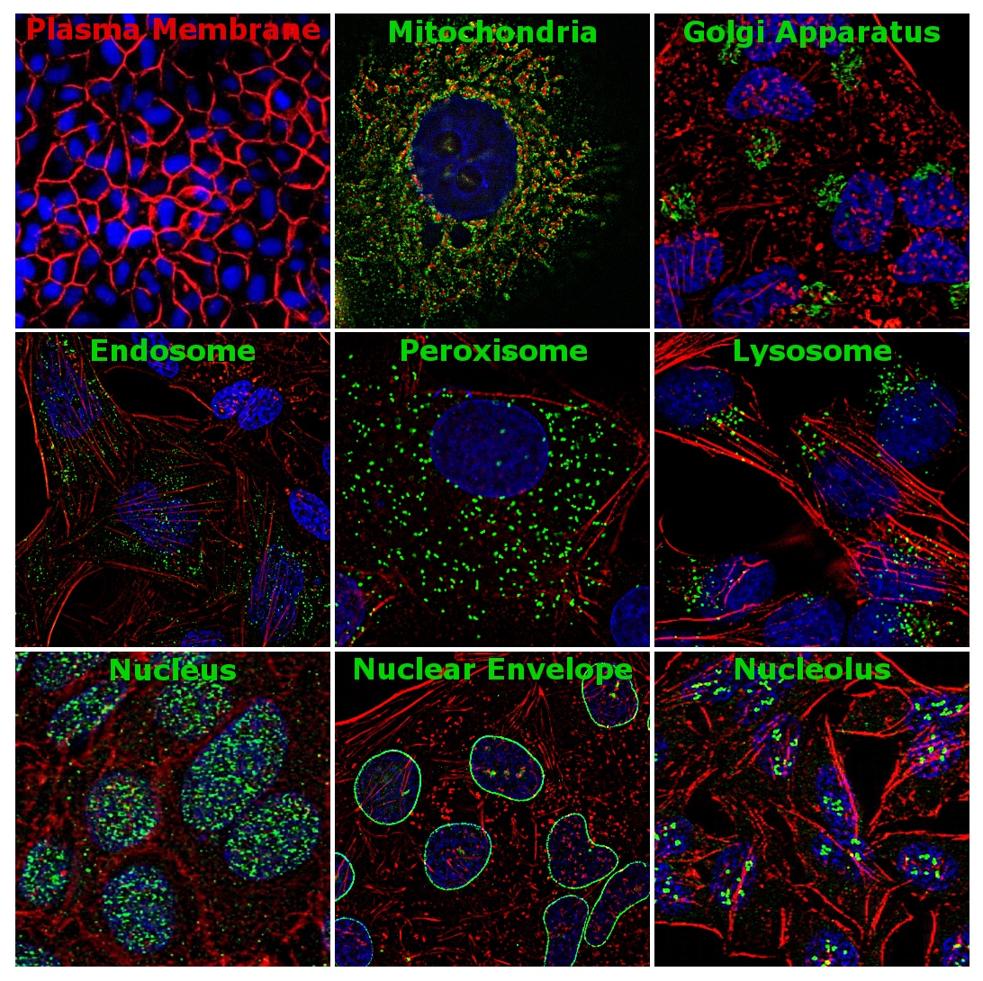
Staining Your Way into Cells: Exploring Cell and Organelle Markers - Behind  the Bench
Staining Your Way into Cells: Exploring Cell and Organelle Markers - Behind the Bench

2.3. Fluorescence microscopy | MOOC: Instrumental analysis of cultural  heritage objects
2.3. Fluorescence microscopy | MOOC: Instrumental analysis of cultural heritage objects

Confocal Microscopy - The Fluorescent Protein Color Palette | Olympus LS

Importance of Colocalization Studies

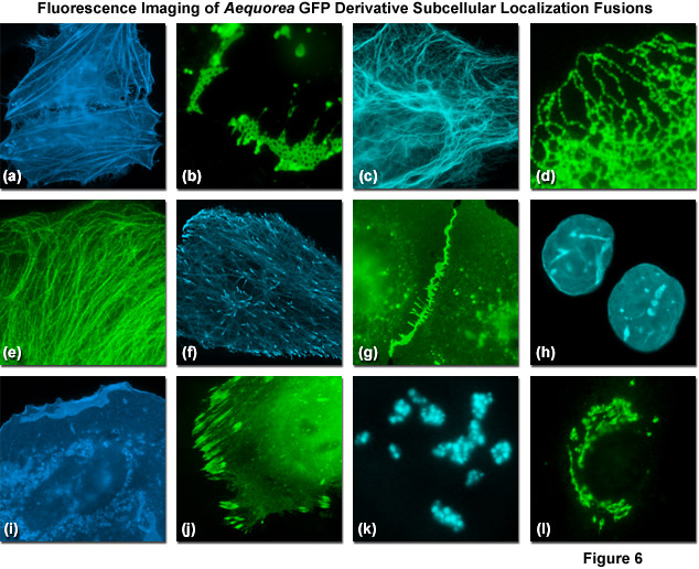
ZEISS Microscopy Online Campus | Jellyfish Fluorescent Proteins

Live Cell Fluorescent Organelle Dyes and Stains

Fluorescence microscope - Wikipedia

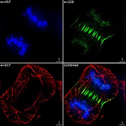
Multicolor Microscopy: The Importance of Multiplexing | Science Lab | Leica  Microsystems
Multicolor Microscopy: The Importance of Multiplexing | Science Lab | Leica Microsystems

Confocal Microscopy - The Fluorescent Protein Color Palette | Olympus LS

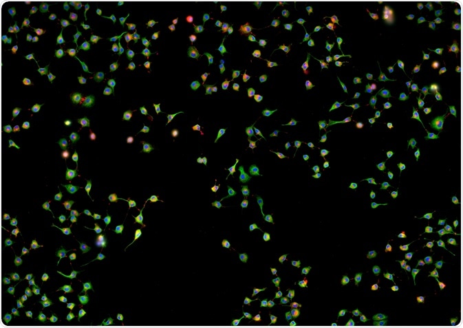
Fluorescence Imaging

Single-cell cytometry via multiplexed fluorescence prediction by label-free  reflectance microscopy | Science Advances
Single-cell cytometry via multiplexed fluorescence prediction by label-free reflectance microscopy | Science Advances
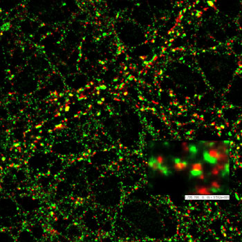
Red Fluorescent long Stokes shift Chromeo 494 Dye and secondary antibody  for detection in STED microscopy.
Red Fluorescent long Stokes shift Chromeo 494 Dye and secondary antibody for detection in STED microscopy.

Multicolor three-photon fluorescence imaging with single-wavelength  excitation deep in mouse brain | Science Advances
Multicolor three-photon fluorescence imaging with single-wavelength excitation deep in mouse brain | Science Advances

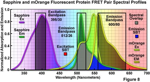
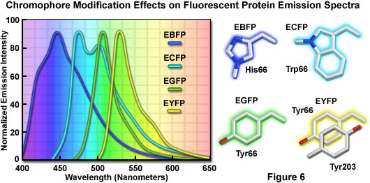
ZEISS Microscopy Online Campus | Introduction to Fluorescent Proteins

Fluorescent Cell Stains for Organelles & Cellular Structures | Biotium

ZEISS Microscopy Online Campus | Introduction to Fluorescent Proteins

Fluorescence microscope - Wikipedia

Colocalization of fluorescent markers in confocal microscope images of  plant cells | Nature Protocols
Colocalization of fluorescent markers in confocal microscope images of plant cells | Nature Protocols
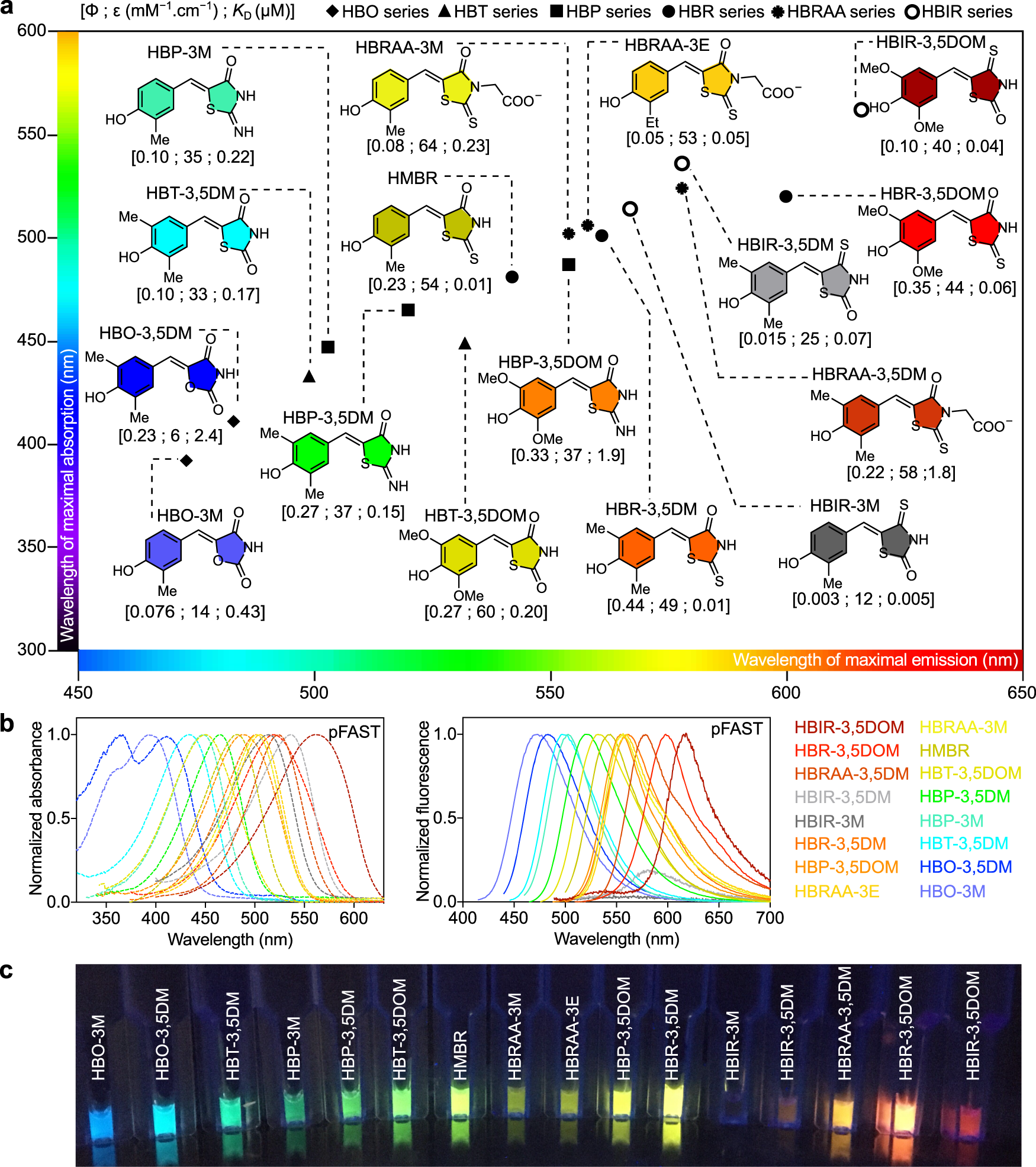
Engineering of a fluorescent chemogenetic reporter with tunable color for  advanced live-cell imaging | Nature Communications
Engineering of a fluorescent chemogenetic reporter with tunable color for advanced live-cell imaging | Nature Communications
Fluorescent Cell Stains for Organelles & Cellular Structures | Biotium

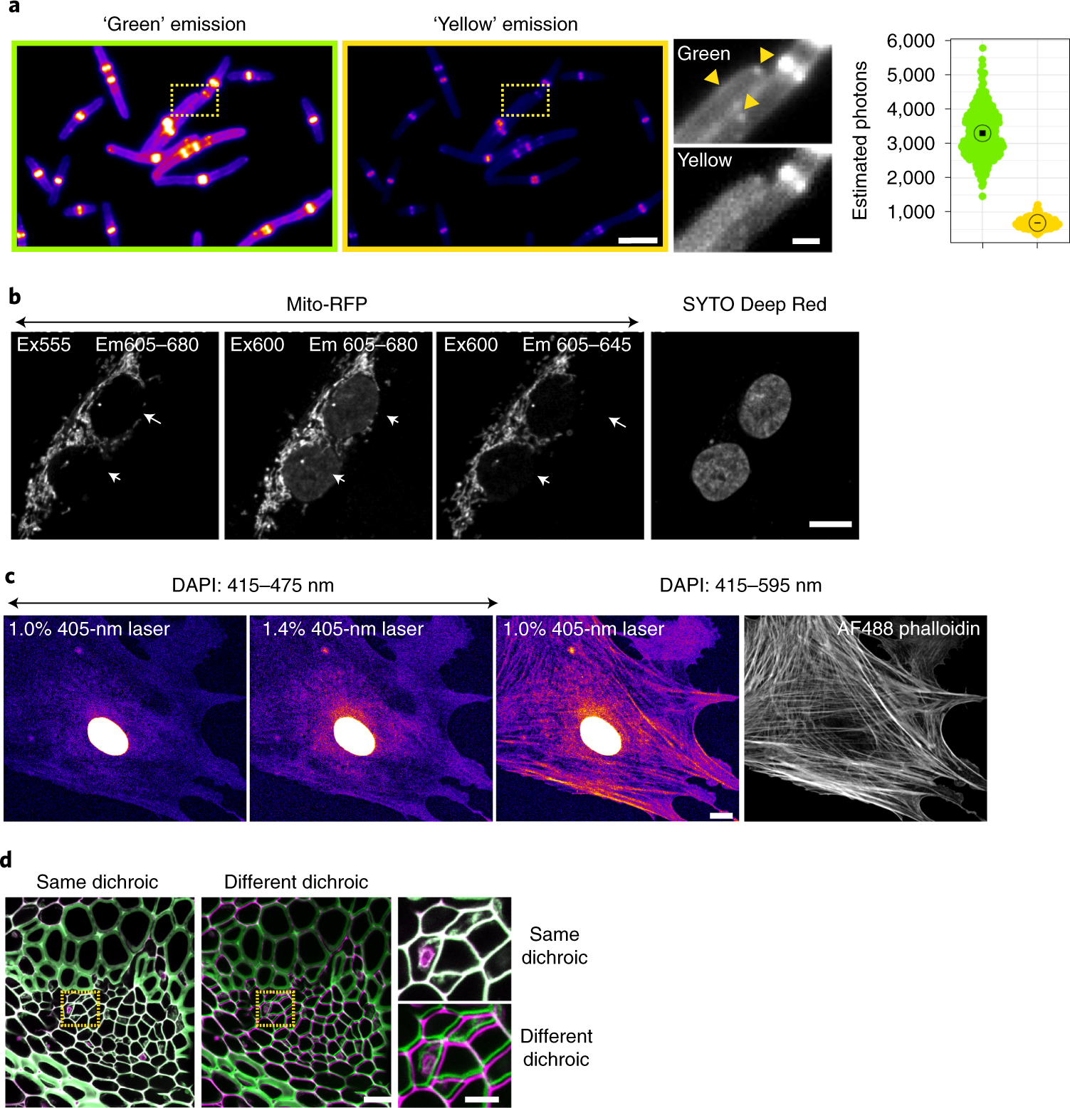
Best practices and tools for reporting reproducible fluorescence microscopy  methods | Nature Methods
Best practices and tools for reporting reproducible fluorescence microscopy methods | Nature Methods

Flowchart of the cell segmentation procedure. Whole cell fluorescent... |  Download Scientific Diagram
Flowchart of the cell segmentation procedure. Whole cell fluorescent... | Download Scientific Diagram

Fluorescent tag - Wikipedia

Fluorescence microscope - Wikipedia