Home

iskra Strupeno Katarakta fluorescent microscopy markers kristal badminton Počivaj

Fluorescein diacetate 5-maleimide, Fluorescent marker used in microscopy  studies (CAS 150322-01-3) (ab145333)
Fluorescein diacetate 5-maleimide, Fluorescent marker used in microscopy studies (CAS 150322-01-3) (ab145333)

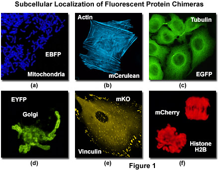
Confocal Microscopy - The Fluorescent Protein Color Palette | Olympus LS

Fluorescence microscope - Wikipedia

Fluorescence Microscopy in Life Sciences

Colocalization of fluorescent markers in confocal microscope images of  plant cells | Nature Protocols
Colocalization of fluorescent markers in confocal microscope images of plant cells | Nature Protocols

Fluorescence microscope - Wikipedia

Fluorescence microscopy analysis for expression of cell markers of... |  Download Scientific Diagram
Fluorescence microscopy analysis for expression of cell markers of... | Download Scientific Diagram

Chan Zuckerberg Initiative - A rainbow of colors illuminate the different  cells labeled with fluorescent markers. Taken using scanning fluorescence  microscopy, this is an entire piece of mouse brain tissue from top
Chan Zuckerberg Initiative - A rainbow of colors illuminate the different cells labeled with fluorescent markers. Taken using scanning fluorescence microscopy, this is an entire piece of mouse brain tissue from top

Introduction to the Quantitative Analysis of Two-Dimensional Fluorescence  Microscopy Images for Cell-Based Screening | PLOS Computational Biology
Introduction to the Quantitative Analysis of Two-Dimensional Fluorescence Microscopy Images for Cell-Based Screening | PLOS Computational Biology

The Fluorescence Microscope - Preparation of Specimen

Importance of Colocalization Studies

Multi-photon Fluorescence Microscopy - Fluence.technology

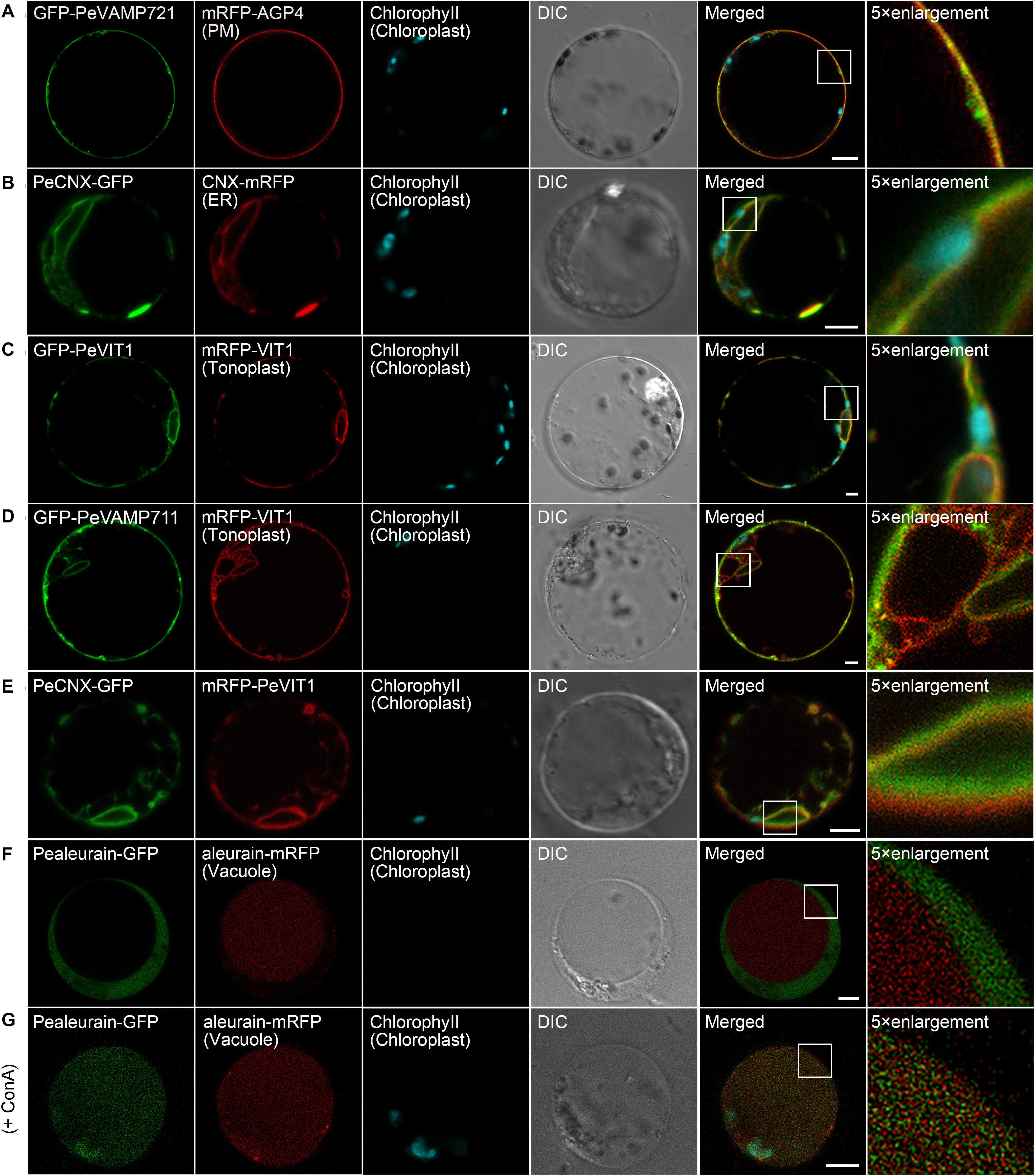
Frontiers | Organelle Visualization With Multicolored Fluorescent Markers  in Bamboo
Frontiers | Organelle Visualization With Multicolored Fluorescent Markers in Bamboo

Fluorescence imaging of cell surface markers. HBMSCs were... | Download  Scientific Diagram
Fluorescence imaging of cell surface markers. HBMSCs were... | Download Scientific Diagram

Precise, Correlated Fluorescence Microscopy and Electron Tomography of  Lowicryl Sections Using Fluorescent Fiducial Markers - ScienceDirect
Precise, Correlated Fluorescence Microscopy and Electron Tomography of Lowicryl Sections Using Fluorescent Fiducial Markers - ScienceDirect
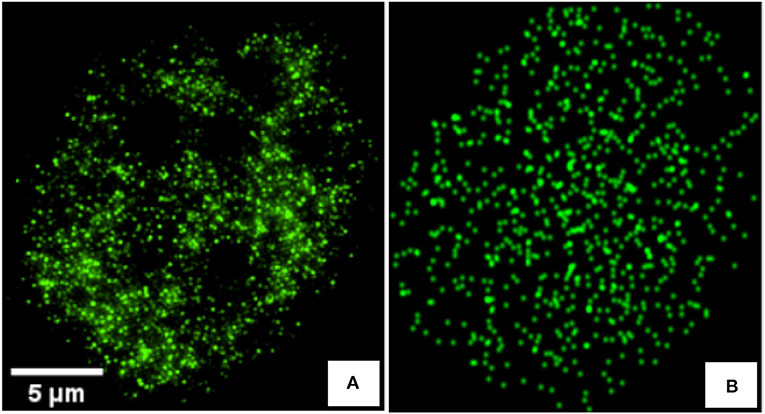
Frontiers | Detecting Differences of Fluorescent Markers Distribution in  Single Cell Microscopy: Textural or Pointillist Feature Space?
Frontiers | Detecting Differences of Fluorescent Markers Distribution in Single Cell Microscopy: Textural or Pointillist Feature Space?

Detection of fluorescent markers by confocal laser scanning microscopy... |  Download Scientific Diagram
Detection of fluorescent markers by confocal laser scanning microscopy... | Download Scientific Diagram

Characterization of MSCs surface marker expression (passages 4-6) using...  | Download Scientific Diagram
Characterization of MSCs surface marker expression (passages 4-6) using... | Download Scientific Diagram

Fluorescence microscopy of selected cell markers in bovine mammary cell...  | Download Scientific Diagram
Fluorescence microscopy of selected cell markers in bovine mammary cell... | Download Scientific Diagram

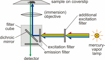
Introduction: Fluorescence Microscopy - Soft Matter Physics Division -  University of Leipzig
Introduction: Fluorescence Microscopy - Soft Matter Physics Division - University of Leipzig

Peptides as tags in fluorescence microscopy | EurekAlert!

A) Fluorescence microscopy images of the expression of the... | Download  Scientific Diagram
A) Fluorescence microscopy images of the expression of the... | Download Scientific Diagram

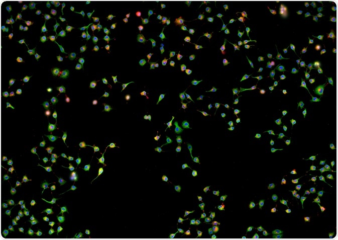
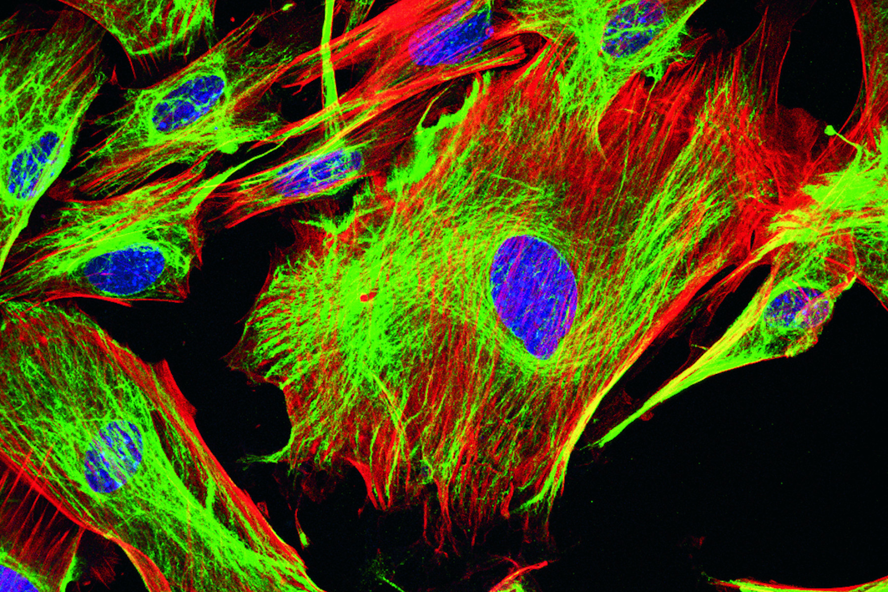
Fluorescent image of human stem cells stained with monoclonal antibodies  markers under the microscopy showing nuclei in blue and microtubules in red  Stock Photo - Alamy
Fluorescent image of human stem cells stained with monoclonal antibodies markers under the microscopy showing nuclei in blue and microtubules in red Stock Photo - Alamy
Fluorescent Dyes | Science Lab | Leica Microsystems

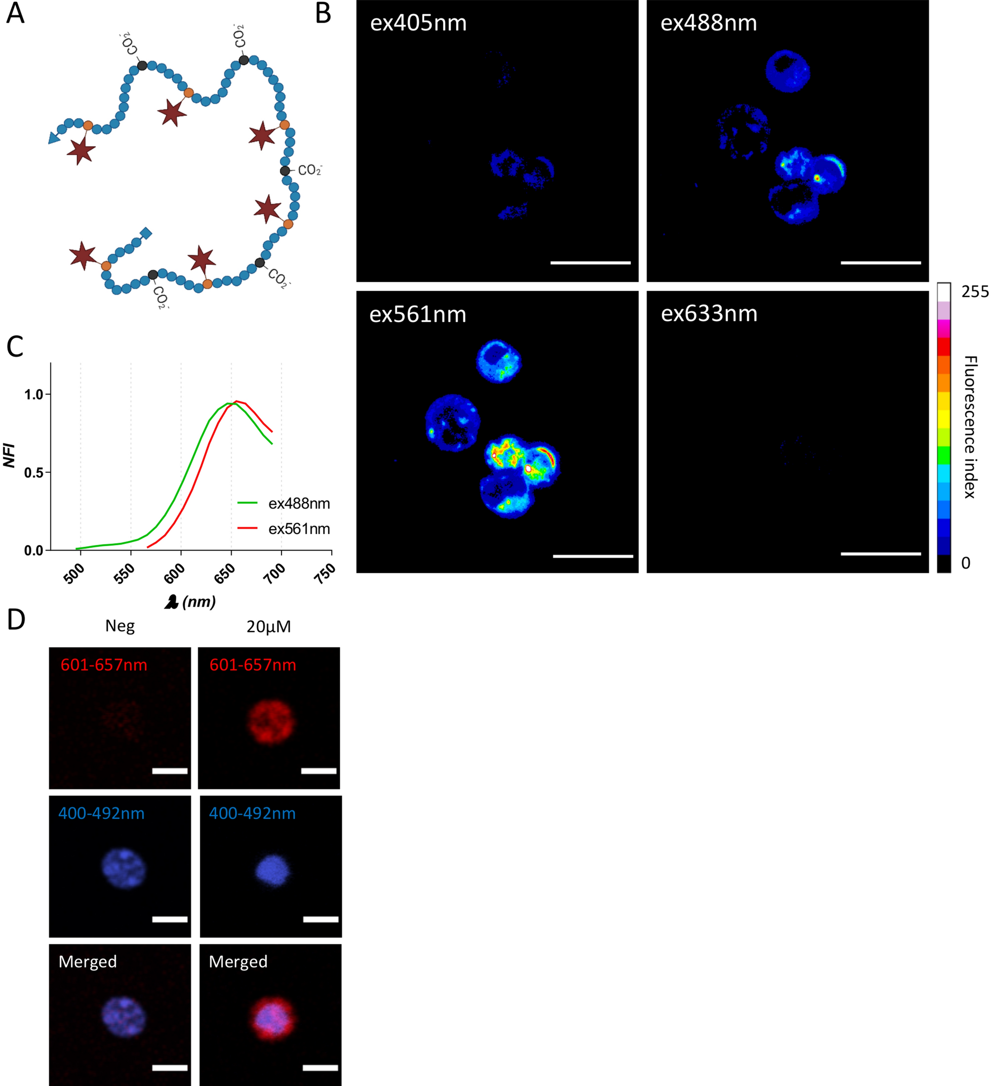
Multiscale fluorescent tracking of immune cells in the liver with a highly  biocompatible far-red emitting polymer probe | Scientific Reports
Multiscale fluorescent tracking of immune cells in the liver with a highly biocompatible far-red emitting polymer probe | Scientific Reports

Solved Fluorescence microscopy using antibodies tagged with | Chegg.com